Synovial Invaginations of the Navicular Bone: What’s Normal, What’s Not?
This article is written for practicing equine veterinarians and reflects specialist-level interpretation of equine foot radiographs in equine diagnostic imaging, incorporating published reference standards, clinical experience, and case-based diagnostic reasoning used in referral and field practice.
You’ve acquired a good-quality dorsoproximal-palmarodistal oblique (DPr-PaDiO) image of the navicular bone. There are six radiolucent zones (synovial invaginations) at the distal border – maybe seven, depending on how you count them.
The horse shows mild, bilateral forelimb lameness. There’s some improvement to palmar digital blocks, but the radiographs don’t show anything else obviously abnormal. So the questions that arise are:
Is this just an incidental finding, or is it evidence of navicular disease or anything else?
Is improvement in lameness rather than abolition of lameness sufficient?
What other structures may be affected by palmar digital nerve blocks?
This is where many veterinarians lose diagnostic clarity. It’s not only about how many synovial invaginations you see. It’s about their shape, size and position , and whether they occur in a horse that is actually lame.
Synovial invaginations are a normal anatomical feature. Most sound horses have them. But without context – or with rushed interpretation, they’re often misread or overlooked entirely.
This article breaks down when invaginations matter, what red flags you should know, and how to interpret them as part of the full clinical picture.
Clinical Decision Summary (For Equine Veterinarians and Veterinary Professionals)
When interpreting synovial invaginations of the navicular bone on DPr-PaDiO radiographs:
- Normal variation is most likely when:
- ≤5–6 triangular invaginations
- Shallow depth (<⅓ of navicular height)
- Sharp margins
- Bilateral symmetry
- Horse is clinically sound
- Further investigation is warranted when:
- Invaginations are widened, irregular, mushroom-shaped or bifid
- Depth exceeds one-third of the bone
- Findings are asymmetric between limbs
- The horse shows lameness responsive to DIP joint or navicular bursa anaesthesia
- Do not interpret in isolation:
- Always correlate with clinical findings, response to diagnostic analgesia, hoof conformation, and additional imaging
- Abnormal synovial invaginations may reflect DIP joint disease, navicular bone pathology, or associated soft tissue injury, rather than primary navicular disease alone
This framework helps prevent both over-diagnosis in sound horses and missed pathology in subtly lame ones.
What Are Synovial Invaginations?
Synovial invaginations are small channels that extend from the distal interphalangeal (distal interphalangeal joint) into the distal border of the navicular bone and are part of the normal anatomical structure.
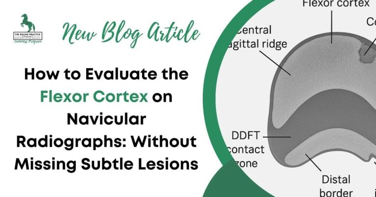
On DPr-PaDiO radiographs synovial invaginations are seen as triangular, semicircular, mushroom-shaped or bifid radiolucent zones along the distal horizontal border of the navicular bone.
In most sound horses, you’ll see a few of these invaginations, mostly triangular in shape, distributed evenly along the distal border of the bone or clustered axially.
They’re not lesions. They’re not inherently abnormal. But in certain contexts, synovial invaginations can indicate early pathology.
Understanding when they are simply part of the bone’s anatomy – and when they suggest a clinical problem – is critical to making the right call in the field.

What Does a Normal Navicular Bone Look Like on Radiographs?
In a sound horse, synovial invaginations are often present. Most veterinarians will see them regularly, especially on well-positioned DPr-PaDiO views.
But not all invaginations are equal – and knowing what’s within normal limits helps prevent both overdiagnosis and missed pathology.
Here’s what typical, non-pathologic invaginations look like:
- Number: Up to five is generally considered normal, especially when evenly spaced and bilateral. Butler, J., Colles, C., Dyson, S. et al. Clinical Radiology of the Horse, 4th ed., 2020, pp. 284–285.
- Shape: Triangular
- Depth: They do not extend more than one-third of the bone’s depth. Most are superficial.
- Margins: Sharply defined.
- Symmetry: Similar appearance between left and right navicular bones increases confidence that they’re incidental.
The presence of a few uniform, symmetrical invaginations – especially in a sound horse with no clinical signs – does not indicate navicular disease.
An increased number of synovial invaginations of variable shape and size may reflect a problem.
However, remember that the navicular bone is an integral part of the distal interphalangeal joint joint, and enlarged synovial invaginations may reflect primary DIP (distal interphalangeal) joint pathology, such as chronic synovitis or osteoarthritis.
This interpretation is supported by CT arthrography work showing that distal border synovial invaginations consistently filled following distal interphalangeal joint arthrography but not following navicular bursa bursography, suggesting these invaginations communicate with the DIP joint and may reflect DIP synovitis/arthropathy rather than primary navicular bone pathology (Olive & Videau, 2017).
Abnormal Synovial Invaginations: Radiographic Red Flags
Not all synovial invaginations are benign. Certain changes in appearance, number, or distribution should prompt closer scrutiny – especially in a horse with clinical signs of lameness.
Here’s what to watch for:
- Excessive number: More than six, especially if they’re clustered or asymmetric.
- Alterations in shape: The presence of semicircular, mushroom-shaped or bifid radiolucent zones along the distal horizontal border of the navicular bone is more likely to be of clinical significance than those of triangular shape.
- Abnormal depth: Invaginations that extend beyond the distal third of the navicular bone.
- Position: Differentiate carefully between radiolucent zones that clearly communicate with the distal border and a circular radiolucent area that does not and which may represent a cyst-like lesion within the bone.
- Position: Radiolucent zones that are present at the junction between the distal horizontal border of the navicular bone and the lateral or medial sloping border usually signify the presence of a distal border fragment within the distal sesamoidean impar ligament. Small fragments are often not detectable on radiographs but can be of clinical significance.
- Asymmetry: If the synovial invaginations look very different in the lame limb compared with the contralateral limb this raises suspicion of DIP joint disease or possibly a form of navicular disease.
- Associated changes: Look for additional radiolucent zones contained within the spongiosa of the navicular bone or in the palmar compact bone; evaluate the shape of the navicular bone in both lateromedial (LM) and DPr-PaDiO images, particularly for evidence of enthesesous new bone at the origin of the distal sesamoidean impar ligament.
- Positive diagnostic blocks: If the horse blocks to the navicular bursa and a large number of synovial invaginations are present of variable shape, they are more likely to be clinically significant. Dyson, S., et al. “Magnetic resonance imaging evaluation of the equine navicular apparatus.” Equine Veterinary Education, 2011.
A large number of synovial invaginations of variable shape don’t confirm navicular disease – they can reflect primary DIP joint disease – or they can reflect previous modelling of the bone and not necessarily progressive on-going pathology.
In many cases, these changes reflect primary distal interphalangeal joint pathology rather than primary disease of the navicular bone itself.
Synovial invaginations are frequently over-interpreted because they are visually striking yet often incidental.
Without correlating limb-specific lameness, response to diagnostic analgesia, and concurrent radiographic changes, there is a risk of incorrectly attributing pain to the navicular bone.
This contributes to both over-diagnosis of navicular disease and under-recognition of primary DIP joint pathology.

Context Is Everything
Synovial invaginations can’t be interpreted in isolation. A radiographic feature is only meaningful when it matches the clinical picture. That’s where many mistakes happen – either overcalling invaginations in a sound horse or dismissing abnormal ones in a horse with clear equine lameness.
Here’s how to bring the full picture into focus:
- History and clinical signs matter. Mild, bilateral forelimb lameness. Palmar foot pain. A positive response to either intra-articular anaesthesia of the DIP joint or a navicular bursa block. These are the signs that should raise your index of suspicion.
- Check the rest of the radiograph. Are there other changes – alterations in shape and size of the entire navicular bone, circular or patchy radiolucent areas within the spongiosa of the navicular bone, palmar compact bone irregularity or focal radiolucent areas, increased opacity of the spongiosa and/or apparent thickening of the trabeculae, reduced demarcation between the palmar compact bone and the spongiosa? In addition carefully evaluate the DIP joint and consider acquisition of dorsolateral-palmaromedial (flexed) and dorsomedial- palmarolateral (flexed) images to better evaluate the articular margins of the middle and distal phalanges. In a LM image carefully assess the distal palmar aspect of the middle phalanx and the dorsoproximal aspect of the navicular bone for the presence of periarticular osteophytes.
- Compare both feet. If the synovial invaginations are asymmetrical, and are more abnormal in the lame limb they are likely to signify other pathology.
- Never ignore the hoof capsule. A long toe, low heel conformation increases mechanical stress on the DIP joint related soft tissues, and the podotrochlear apparatus and the laminar attachments to the distal phalanx.
And if you’re still unsure – get help. That’s what second opinions, repeat radiographs, or referral imaging are for. Uncertainty doesn’t mean indecision. It means doing what’s best for the horse.
Clinical Scenarios – When Invaginations Matter
When synovial invaginations appear abnormal, context guides action. Below are two contrasting real-world cases to help frame clinical judgment:

Case 1: Clinically Significant
Horse: 10-year-old warmblood gelding, upper-level dressage
History: Gradual onset right forelimb lameness, worsening under saddle
Diagnostics:
- Positive response to navicular bursa block
- On a DPr-PaDiO radiograph there were seven synovial invaginations in the distal horizontal border of the right navicular bone
- The synovial invaginations were widened, irregular in shape, and extended more than one-third of the proximodistal height of the navicular bone
- There were areas of increased opacity immediately proximal to/ around the synovial invaginations.
Follow-up: Magnetic resonance imaging (MRI) identified a partial-thickness tear of the deep digital flexor tendon and increased signal intensity in the palmar third of the navicular bone from proximal to distal in fat suppressed images, which could reflect oedema, haemorrhage, fibrosis or bone necrosis.
Outcome: Diagnosis of combined pathology of the deep digital flexor tendon and the navicular bone. Follow-up MRI after rest will often demonstrate resolution of the increased signal intensity in the palmar third of the navicular bone in fat suppressed images which develops in association with DDFT lesions.
In this case, the synovial invaginations were not normal and may reflect either concurrent DIP joint disease or the potential for progressive navicular bone pathology.

Case 2: Incidental Finding
Horse: 7-year-old off-the-track Thoroughbred, in light paddock work
History: No history of lameness
Diagnostics:
- on routine prepurchase radiographs there were six synovial invaginations in each navicular bone
- The synovial invaginations were triangular, small and narrow, were symmetric, and were confined to the distal border of the navicular bone
- No clinical signs of lameness
- The front feet clearly had low-heel conformation typical of many Thoroughbreds and careful trimming and shoeing is important
Follow-up: No specific treatment needed, monitored over 12 months, remained sound
In this case, the synovial invaginations were normal variation. No intervention was required.
These scenarios reinforce a simple truth: synovial invaginations are only part of the picture. What matters is how they relate to the horse in front of you.
Final Takeaways for the Field Veterinarian
Synovial invaginations are not a diagnosis. They are a feature – sometimes normal, sometimes not. What determines their relevance is how they appear, how many there are, and whether they match the clinical picture.
Here’s what to remember:
- Don’t count alone. The number of invaginations is less important than their size, shape, symmetry, and depth.
- Look for red flags. Widened, irregular, or deep invaginations – especially when asymmetric or accompanied by changes in the architecture of the spongiosa – warrant further attention.
- The navicular bone is part of the DIP joint: Assess the distal interphalangeal joint carefully and consider acquiring oblique (flexed) images to better evaluate the joint margins.
- Match findings to the horse. Is the horse lame? Does it improve to intra-articular anaesthesia of the DIP joint or a navicular bursa block? Are there conformational factors that add mechanical stress to the palmar aspect of the foot?
- Always compare both feet. Asymmetry often tips you off.
- When in doubt, repeat or acquire additional radiographic projections, ask for help with interpretation or refer. A sufficient number of images of the appropriate structures and a fresh set of eyes can make all the difference.
Synovial invaginations along the distal horizontal border of the navicular bone or at the junction between the horizontal and medial or lateral sloping borders of the bone can signify clinical disease of either the distal interphalangeal joint or the navicular bone.
Prognostic Insight
The prognostic weight of synovial invaginations depends on their radiographic features, the clinical context, and any associated findings:
- Up to six small triangular shaped radiolucent zones along the distal horizontal border of the navicular bone is commonly seen in normal horses.
- A large lucent zone at the junction of the distal horizontal and medial and/ or lateral sloping border is likely to be associated with a distal border fragment of the navicular bone which can cause pain and lameness. Large fragments can be identified radiographically but small fragments often cannot.
- Variably shaped and sized synovial invaginations can reflect primary DIP joint disease or may be an indicator of some form of navicular bone pathology.
This framework can help you decide when to act, when to watch, and when to refer.