ECVM in Horses? What C6-C7 Variations Mean on Radiographs
Study reviewed: Dyson S, Phillips K, Zheng S and Aleman M. Congenital variants of the ventral laminae of the sixth and seventh cervical vertebrae are not associated with clinical signs or other radiological abnormalities of the cervicothoracic region in Warmblood horses. Published online on 28 June 2024 and in Equine Veterinary Journal, March 2025;57(2):419-430. DOI: 10.1111/evj.14127
ECVM, or equine complex vertebral malformation, is a term frequently used in online searches and owner discussions about congenital variations of the caudal cervical vertebrae. However, it is not the preferred scientific terminology in this article. The word “malformation” may imply pathology before the clinical significance of the finding has been established.
Dyson et al. also cautioned against equating these equine variations with complex vertebral malformation syndrome in Holstein cattle. The conditions are not equivalent.
In this article, I use the more neutral terms congenital C6-C7 variation and equine caudal cervical morphologic variation, or ECCMV. These variations may include the absence of one or both ventral laminae from C6, sometimes with corresponding bone on the ventral aspect of C7.
Dr Sue Dyson and colleagues investigated whether congenital C6-C7 variants were associated with clinical signs or other radiological abnormalities in a population of Warmblood horses.

The Study Design
This cross-sectional study examined 223 Warmbloods aged ≥3 years seen at two referral centres:
- 96 cases with neurologic signs, neck pain/stiffness, or neck-related forelimb lameness, and
- 127 controls with no clinical signs referrable to the neck.
Each horse underwent a standardised orthopaedic and neurologic assessment and high-quality radiographs (C5 → T2). Images were reviewed blind to case status using a pre-defined grading system.
The 223-Horse Warmblood Study
This cross-sectional study examined 223 Warmblood horses aged three years or older at two referral institutions:
- 96 clinical cases with neurologic signs, neck pain or stiffness, or neck-related forelimb lameness
- 127 controls without clinical signs referable to the neck
All horses underwent standardised orthopaedic and neurologic assessments.
The radiographic examination included laterolateral and paired left and right 45-55 degree ventrolateral-dorsolateral oblique views from C5 to T2. At each institution, the images were assessed by one reader who was blinded to the horses’ clinical categories, using a predefined grading system.
Key Findings
- The ventral profile of C7 was abnormal in 54 of the 223 horses, or 24.2%.
- Congenital variants of C7 were identified in 16.7% of the clinical cases and 29.2% of the controls.
- The presence of a congenital C7 variant was not associated with the type of clinical case or with the other radiological findings assessed in the cervicothoracic region.
- There was no association between the congenital variants and articular-process modelling, discospondylosis, spondylolisthesis or intervertebral-foramen narrowing.
- Severe articular-process modelling at C6-C7 and C7-T1 was more frequent among clinical cases than controls. These findings were separate from the presence or absence of a congenital C6-C7 variation.
Interpretation
In this Warmblood population, congenital C6-C7 variants were common and were not associated with the clinical case categories investigated. The findings support interpreting the variation in clinical context rather than treating its presence alone as a diagnosis.
It is difficult to explain how congenital variants, in their own right, could cause clinical signs unless secondary osseous or soft-tissue changes resulted in spinal-cord compression, nerve-root impingement or pain.
Although these variations may alter local anatomy and longus colli muscle attachments, this study did not demonstrate an association with clinically significant disease in the Warmblood population examined.
This does not mean that every caudal cervical radiographic finding is benign. Severe articular-process modelling, malalignment, spondylolisthesis, discospondylosis and other abnormalities must be assessed separately and interpreted alongside the horse’s clinical examination.
The study was cross-sectional and included Warmblood horses only. The radiographs were read independently by one assessor at each institution. Its conclusions should not be applied without qualification to every breed or clinical population.
This reinforces why radiographic findings must match the horse rather than being interpreted in isolation.
What the 2026 Pre-Purchase Study Adds
A 2026 retrospective study reviewed 200 clinically sound Warmblood sport horses presented for pre-purchase examinations at an equine hospital in Germany between 2020 and 2024. The cervical radiographs assessed in the study had been obtained as part of those pre-purchase examinations.
Equine caudal cervical morphologic variation, or ECCMV, was identified in 30% of the horses. C6-C7 articular-process joint modelling was identified in 32.5%.
The researchers found no significant association between ECCMV and articular-process joint modelling. Recorded competition results also did not differ significantly between horses with and without ECCMV.
These findings provide additional evidence that this congenital variation can be identified in clinically sound Warmblood sport horses without an observed reduction in the competition outcomes measured by the study. However, the study was retrospective and was not a longitudinal assessment of future soundness. Its results cannot guarantee the future soundness or performance of an individual horse.
Read the 2026 Warmblood pre-purchase and competition study.

Interpreting the 2024 JAVMA Study
A separate 2024 retrospective observational study reviewed the records of 135 horses that had undergone cervical radiography. Transposition of the ventral lamina from C6 to C7 was more prevalent in a small subgroup of horses with neck pain than in horses without neck pain.
However, this finding demonstrated an association only. It did not establish that the congenital variation caused the neck pain.
The study found no differences in the final diagnoses of neurologic disease, cervical orthopaedic disease or lameness between horses with and without the variation. Its retrospective design and the small neck-pain subgroup are important methodological limitations. The authors recommended further standardised prospective research.
Read the 2024 JAVMA study.
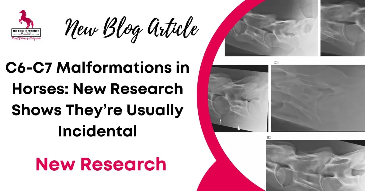
What C6-C7 Variations Look Like on Radiographs
Recognised radiographic appearances may include:
- Unilateral or bilateral absence of the ventral laminae from C6
- A ventral lamina or vestigial process appearing on C7
- Unilateral or bilateral transposition
- Asymmetry between the left and right sides
A laterolateral view may demonstrate the ventral profiles of C6 and C7. Paired oblique views can assist with assessing sidedness and the articular processes.
The 223-horse study assessed the region from C5 to T2 rather than considering C6 and C7 in isolation. Radiographs describe the osseous morphology, but they do not, by themselves, identify a source of pain or establish a neurologic diagnosis.
For broader context on interpreting common imaging findings, read When Radiographic Abnormalities Are Common in Performance Horses.
Clinical Take-Home Messages
- ECVM is a commonly searched but disputed term. Congenital C6-C7 variation or ECCMV provides more neutral anatomical terminology.
- A congenital C6-C7 variation on radiographs does not, by itself, establish the source of neck pain, stiffness, lameness or neurologic dysfunction.
- In the 223-horse Warmblood study, the congenital variation was identified more frequently among controls than clinical cases.
- A congenital variation in its own right is unlikely to cause clinical signs unless secondary osseous or soft-tissue changes result in pain, nerve-root impingement or spinal-cord compression.
- Severe articular-process modelling and other caudal cervical abnormalities should be assessed separately from the congenital variation.
- The 2026 study found no significant difference in recorded competition outcomes between clinically sound Warmbloods with and without ECCMV. These horses had undergone radiography as part of pre-purchase examinations.
- Radiographs must be interpreted alongside the horse’s history, orthopaedic examination, neurologic examination, intended use and any additional imaging findings.
How to Explain the Finding to an Owner or Buyer
The radiographs show a congenital variation in the shape of the lower cervical vertebrae. This type of finding is also seen in clinically normal horses. On its own, it does not diagnose or predict pain, neurologic disease or future performance problems.
In the absence of clinical features related to the neck or other radiographic abnormalities, the finding is unlikely to be clinically significant.
No pre-purchase examination can accurately predict the future. Judgement must be based on what is observed on the day, including the horse’s history, clinical examination, intended use and any accompanying abnormalities.
If a practitioner is uncertain about the radiological interpretation, the radiographs should be sent to an expert for a second opinion before making a final recommendation regarding purchase.
Frequently Asked Questions About ECVM and C6-C7 Variations
What is ECVM in horses?
ECVM, or equine complex vertebral malformation, is a commonly used but disputed term for congenital variations affecting the caudal cervical region, particularly C6 and C7. The term does not establish that the anatomical variation is causing disease in an individual horse. This article uses the more neutral terms congenital C6-C7 variation and ECCMV.
Are C6-C7 malformations the same as ECVM?
C6-C7 variations are among the findings commonly discussed under the ECVM label. However, definitions and terminology vary. The specific anatomical finding should be described rather than relying on the ECVM label alone.
Is ECVM the same as wobbler syndrome?
No. Wobbler syndrome generally refers to cervical vertebral compressive myelopathy. A congenital variation of the ventral laminae of C6 and C7 does not, by itself, diagnose spinal-cord compression or neurologic disease.
What symptoms does ECVM cause?
There is no reliable list of clinical signs that proves a congenital C6-C7 variation is the cause. Neck pain, stiffness, forelimb lameness, poor performance and neurologic deficits may warrant investigation, but they should not automatically be attributed to the radiographic variation.
A causal explanation would require consideration of secondary osseous or soft-tissue changes that could result in pain, nerve-root impingement or spinal-cord compression.
Can a horse with a C6-C7 variation compete normally?
Yes, these variations have been documented in clinically sound Warmblood sport horses. In the outcomes measured by the 2026 study, competition results did not differ significantly between horses with and without ECCMV.
The horses were radiographed as part of pre-purchase examinations. This population-level finding does not guarantee the future soundness or performance of an individual horse.
Should a C6-C7 variation fail a pre-purchase examination?
Not automatically. Its significance depends on the horse’s history, clinical findings, intended use, other imaging findings and the buyer’s risk tolerance.
No pre-purchase examination can accurately predict the future. If the practitioner is uncertain about the radiological interpretation, the images should be referred to an expert for a second opinion before making a final recommendation regarding purchase.
For further clinical education on lameness assessment and diagnostic imaging, explore our equine lameness continuing education resources and the Equine Lameness Program.
Related Research and Sources
Two related papers from the same 223-horse population provide further information about the clinical picture associated with caudal neck pathology and the other radiological abnormalities identified in the cervicothoracic region.
These papers address separate questions from the congenital C6-C7 variation and should be considered alongside the principal study.
- Dyson S, Phillips K, Zheng S and Aleman M. Congenital variants of the ventral laminae of the sixth and seventh cervical vertebrae are not associated with clinical signs or other radiological abnormalities of the cervicothoracic region in Warmblood horses. Equine Veterinary Journal. 2025;57(2):419-430. DOI: 10.1111/evj.14127
- Dyson S, Zheng S and Aleman M. Primary phenotypic features associated with caudal neck pathology in Warmblood horses. Journal of Veterinary Internal Medicine. 2024;38(4):2380-2390. DOI: 10.1111/jvim.17125
- Dyson S, Quiney L, Phillips K, Zheng S and Aleman M. Radiological abnormalities of the cervicothoracic vertebrae in Warmblood horses with primary neck-related clinical signs versus controls. Veterinary Radiology & Ultrasound. 2024;65:755-768. DOI: 10.1111/vru.13420
- Henderson CS, Story MR and Nout-Lomas YS. Neck pain but not neurologic disease occurs more frequently in horses with transposition of the ventral lamina from C6 to C7. Journal of the American Veterinary Medical Association. 2024;262(9):1215-1221. DOI: 10.2460/javma.24.04.0230
- Strootmann T, Peter VG and Körner J. Radiographic Prevalence of Anatomical Variations of the Ventral Lamina of the Sixth Cervical Vertebra, C6/C7 Articular Process Joint Modelling and Competition Outcomes in Warmblood Sport Horses. Animals. 2026;16(3):424. DOI: 10.3390/ani16030424
Read the Full Open-Access Paper
You can explore the complete paper including the methodology, tables, and image examples below.
© 2024 The Authors. Published in Equine Veterinary Journal by John Wiley & Sons Ltd under the Creative Commons Attribution-NonCommercial-NoDerivs License CC BY-NC-ND 4.0. DOI: 10.1111/evj.14127