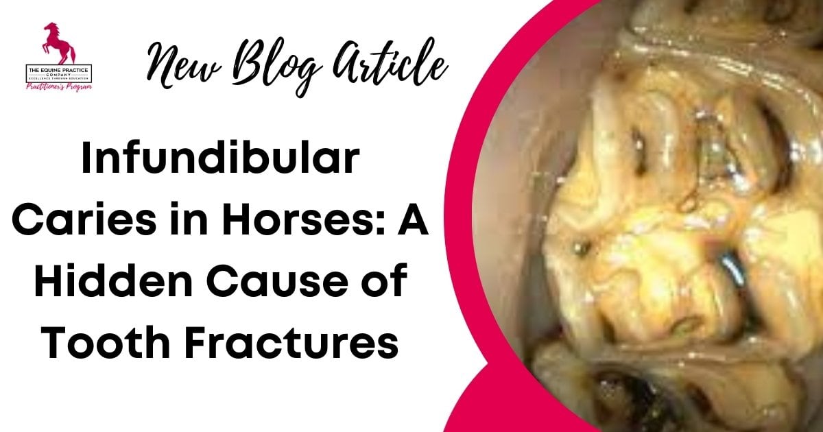
Infundibular Caries in Horses: Early Diagnosis, Grading, and Fracture Risk
You’ve got the horse sedated, the speculum in place, and your mirror positioned. There’s something off about the 109s. A dark, irregular patch inside the infundibulum. Is it cemental wear? A feed stain? Early caries?
You keep going. The horse finishes the float. No signs of pain, no swelling, nothing urgent.
Six months later, the same horse presents with a fractured tooth and a unilateral nasal discharge. Radiographs confirm what you’d suspected too late: a sagittal fracture through a carious infundibulum.
The fracture could’ve been prevented, if the lesion had been recognised and staged earlier.
This is the quiet risk of infundibular caries. It doesn’t always look like much. But once the crown structure fails, you’re no longer treating a defect. You’re now managing a surgical case.
And that’s why it matters.
Quick Clinical Summary (For Equine Veterinarians)
Infundibular caries is a common, often under-recognised cause of maxillary cheek tooth fracture in horses.
Key points:
- Most commonly affects the 09s–11s
- Early lesions may appear clinically insignificant
- Stage 3-4 lesions significantly weaken crown integrity
- Imaging is essential for accurate staging and treatment planning
- Early recognition can prevent fracture and surgical extraction
In this article, we’ll walk through what infundibular caries actually is, how it contributes to fracture, and the techniques every equine veterinarian needs to know to detect it early in equine dental education.
We’ll also reference Module 2 of the Equine Dentistry Program, where Dr. Kirsten Jackson explains how to stage, image, and treat these cases in practice.

What Is Infundibular Caries?
Infundibular caries is a progressive, destructive process that affects the infundibula of maxillary cheek teeth, most commonly the 09s–11s.
It’s not the same as peripheral caries, which affect the outer cementum and enamel. Infundibular caries begins deep within the tooth and is often hidden during routine oral exams—until it’s too late.
The underlying problem is usually cemental hypoplasia or necrosis. Horses are born with infundibula that may be partially unfilled or filled with under-mineralised cementum.
Over time, these defects can trap feed material and become colonised by acid-producing bacteria. The acidic environment breaks down the cementum and eventually the underlying enamel and dentine.
Once the lesion reaches Stage 4, the structural integrity of the crown is compromised. If left untreated, the lesion becomes a fracture line waiting for the right amount of force to finish the job.
If you need more information, Dr. Kirsten Jackson walks through the classification and progression of infundibular caries in detail in Module 2 of the Equine Dentistry Program, using real clinical cases and radiographs.
She explains how to differentiate mild cemental defects from deeper, fracture-prone caries—and what to do when you spot them.
How Infundibular Caries Is Graded in Horses
Several grading systems have been proposed for infundibular caries in horses, but most classify lesions based on depth, tissue involvement, and structural compromise.
A simplified clinical framework:
Stage 1 – Cemental defects without cavitation
Stage 2 – Cemental loss with early cavitation
Stage 3 – Extension into dentine, structural weakening
Stage 4 – Pulp involvement or complete infundibular collapse
Fracture risk increases substantially once lesions progress beyond Stage 3, particularly in maxillary cheek teeth with bilateral infundibular involvement.
Accurate grading requires sedation, thorough oral examination, and imaging.
Why It Matters: The Link to Tooth Fracture
Once infundibular caries progresses past Stage 3, the risk of fracture starts to rise—especially in the maxillary cheek teeth, where the two infundibula run side-by-side.
As the cementum and dentine break down, the crown loses its internal support. With every chew, the weakened structure absorbs less force and begins to flex. Over time, the result is often a sagittal midline fracture, right through the affected infundibulum.
These fractures aren’t subtle. They often present with:
- Sudden onset oral pain
- Unilateral nasal discharge (if there’s secondary apical infection or sinus involvement)
- Loss of crown structure or displaced fragments
- Abnormal odour or feed impaction
But by this point, your treatment options are limited. In most cases, you’re facing a surgical extraction of a compromised tooth with secondary endodontic disease.
Many of these cases had warning signs – feed packing, visible cavitation, cemental collapse – but they weren’t recognised or investigated early enough. And once the fracture occurs, the damage is permanent.
The takeaway: if you’re seeing infundibular defects during an oral exam, you need to treat them as potential structural failures in progress – not just cosmetic findings.
Recognition and management of infundibular caries is widely discussed in equine dental literature and continuing education, with consensus that advanced lesions are structurally significant rather than cosmetic findings.
Current recommendations emphasise early detection, appropriate imaging, and case-by-case decision-making to reduce fracture risk and avoid secondary apical disease.
How to Diagnose It – Before It’s Too Late
Infundibular caries isn’t hard to diagnose, if you’re looking for it and using the right tools. But too often, it’s missed during rushed or incomplete oral exams.
Here’s what your diagnostic workflow should include:
Oral Examination
Sedation is non-negotiable. You need a still horse and time to assess.
Use a full-mouth speculum, a bright light, and a dental mirror or oral endoscope.
Examine all maxillary cheek teeth, particularly the 09s to 11s.
Look for:
- Feed packing
- Irregular contours or central cavities
- Inappropriate discolouration of the enamel and dentine on the occlusal surface
These findings alone aren’t enough to make a treatment decision – but they should always lead to imaging.
Radiographs and Advanced Imaging
Standard skull radiographs can show changes in mineral density or shadowing, but they’re often limited in sensitivity.
Oblique views (e.g. lateral oblique or extraoral DV) can help visualise apical changes.
If available, standing CT is the gold standard. It allows you to evaluate:
- The depth and extent of the lesion
- Whether one or both infundibula are affected
- Communication with the pulp or apical structures
CT findings also help you differentiate a restorable tooth from one at high risk of fracture or infection.
In Module 2 of the Equine Dentistry Program, Dr. Kirsten Jackson walks through these diagnostic steps in real clinical cases, showing how to combine exam findings, radiographs, and CT to build a complete picture.
She also explains the staging system used in published studies, so you can apply consistent terminology in your notes and referrals.

Treatment Options and Decision-Making
Once you’ve identified infundibular caries, the next question is whether to monitor, restore, or extract. The right approach depends on the stage of the lesion, the integrity of the remaining crown, and whether the pulp or adjacent structures are involved.
When Monitoring Is Appropriate
- Small, shallow cemental defects with no evidence of cavitation
- Staging suggests Stage 1 or early Stage 2
- No signs of infection, feed packing, or pain
- No evidence of structural loss on imaging
These cases should be recorded carefully, re-examined at 6 to 12 month intervals, and radiographed if any progression is suspected.
When to Consider Restoration
Restoration is worth considering when:
- There’s clear cavitation, but no pulp involvement
- The lesion is classified as Stage 2 or 3 and has a probing depth of greater than 25mm
- Both infundibula are affected (bilateral lesions carry higher risk)
- You have access to appropriate materials (e.g. dual-cure composites, glass ionomer)
- The case can be referred to a veterinarian trained in restorative techniques
Restoration can significantly reduce the risk of fracture, especially in younger horses with active masticatory loading.
When Extraction Is Necessary
Extraction should be considered when:
- There is Stage 4 disease with exposure of the pulp
- There is a midline sagittal fracture
- There’s clinical evidence of apical infection or sinusitis
- CT shows loss of crown integrity or significant loss of reserve crown
- The tooth is non-restorable and poses an ongoing risk of pain or pathology
While referral for advanced imaging and treatment is ideal, general practitioners should feel confident recognising these patterns and discussing prognosis and options with the owner early.
Key Takeaways for Busy Practitioners
Infundibular caries is common, under diagnosed, and structurally significant.
- Most fractures of maxillary cheek teeth started with a carious infundibulum that was either missed, underestimated, or compounded by over-floating in equine dentistry.
A proper oral exam matters.
- You can’t assess infundibular health without sedation, good lighting, a mirror or endoscope, and time.
Use imaging to guide your decisions.
- CT is ideal, but even radiographs can show early signs of cavitation or apical change. Don’t skip this step if something looks off.
Not every defect needs a filling – but not every one can wait.
- Monitoring is fine for Stage 1 and 2 lesions. Once dentine or pulp is involved, it’s time to plan for restoration or extraction.
Your notes should reflect staging and risk.
- A clear record helps with rechecks, referrals, and client conversations.
This is preventable pathology.
- Recognising infundibular caries before the fracture means saving the tooth – and avoiding surgical extractions.
Want to See These Cases in Action?
Dr. Kirsten Jackson takes you step-by-step through real-world cases of infundibular caries as part of our advanced equine veterinary dentistry training.
You’ll see how she stages each lesion, interprets CT findings, and makes decisions about restoration versus extraction – using radiographs, intraoral images, and video from live patients.
If you’re already seeing feed packing or irregular infundibula in practice, this module will help you make more confident decisions.
And if you’ve ever had a surprise fractured due to infundibular caries… you’ll wish you’d watched it sooner.
FAQ: Infundibular Caries in Horses
What is infundibular caries in horses?
Infundibular caries is a progressive loss of cementum, enamel, and dentine within the infundibula of maxillary cheek teeth, most commonly the 09s–11s.
Can infundibular caries cause tooth fracture?
Yes. Advanced infundibular caries weakens the internal structure of the crown and is a well-recognised predisposing factor for sagittal midline fractures.
How is infundibular caries graded in horses?
Grading is based on lesion depth and tissue involvement, ranging from cemental defects (Stage 1) to pulp exposure and structural collapse (Stage 4).
Can infundibular caries be treated?
Early-stage lesions may be monitored. Stage 2–3 lesions may be suitable for restorative treatment. Stage 4 disease often requires extraction.
How is infundibular caries diagnosed?
Diagnosis involves sedated oral examination and imaging. Standing CT provides the most accurate assessment of lesion depth and fracture risk.