Equine Periodontal Disease: The Hidden Cause of Tooth Loss, Pain, and Occlusal Collapse
You’ve probably seen it before.
A horse with no obvious pathology, no fractures, no feed packing… and yet a cheek tooth is unexpectedly mobile. Or worse – already lost.
We tend to blame the tooth. But often, the real problem started much earlier, buried in the periodontium – specifically, in the periodontal ligament.
Equine periodontal disease is one of the most common yet overlooked and under-diagnosed causes of progressive dental dysfunction.
Equine periodontal disease refers to inflammation and progressive loss of the periodontal ligament and surrounding alveolar bone, most commonly affecting the cheek teeth.
Early disease may present with subtle sulcal deepening or feed trapping, while advanced cases result in attachment loss, tooth mobility, and eventual tooth loss.
The periodontal ligament plays a critical role in occlusal stability and shock absorption in the hypsodont dentition.
Failure to detect and manage periodontal disease early can lead to irreversible functional collapse of the dental arcade.
And by the time it shows up as mobility, the damage has usually been going on for months, sometimes years.
Equine periodontal disease is one of the most common diseases affecting horses, with published studies and professional veterinary bodies reporting prevalence rates of up to 50%, particularly in ageing populations.
The ligament doesn’t just hold the tooth in place. It:
- Absorbs shock with every chew
- Maintains occlusal stability
- Adapts to the changing forces across the arcade
- And in horses, it becomes more dense and fibrous with age—making it more resilient and, if inflamed, a major source of long-term instability (fun fact: this is why is it easier to extract the tooth of a 4 year old compared with the same tooth of a 12 year old!)
Once inflamed, the cascade begins: feed trapping, endotoxin release, loss of attachment, early mobility, and secondary occlusal changes that can affect the entire mouth and chewing ability.
Most of this is missed during routine exams UNLESS the horse is still (this means sedation), the speculum is placed correctly, and the practitioner is using a strong focal light AND a mirror, AND most importantly, knows what they are looking at.
In this article, we’ll break down:
- What equine periodontal disease actually is
- How it quietly disrupts the entire dental system
- Why it’s missed in many dental appointments
- And how you can detect it early, intervene sooner, and preserve teeth that would otherwise be lost
This isn’t just about doing better equine dentistry. It’s about protecting long term function and comfort.
Let’s start where it all begins: the ligament.

What Is Equine Periodontal Disease And Why Is It So Often Missed?
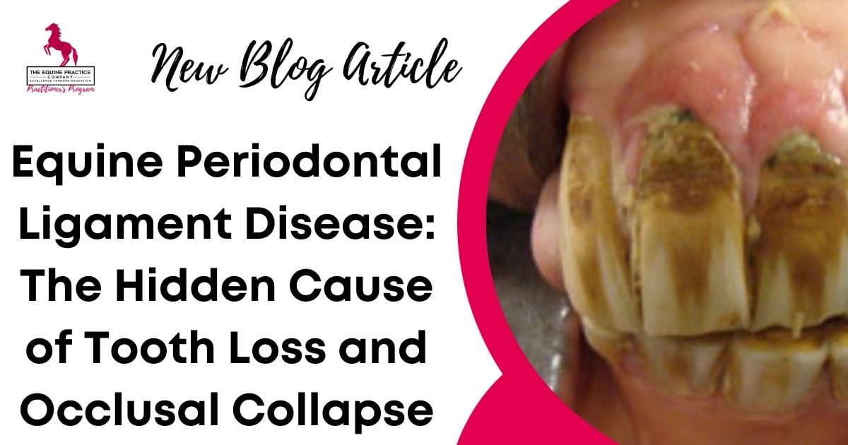
When most equine vets think of periodontal disease, they picture obvious signs such as feed packing, gingival recession, diastemata, and halitosis. But by the time these signs are visible, the damage to the periodontium is usually well advanced.
The periodontal ligament (PDL) is the soft tissue structure that connects the cementum of the tooth to the surrounding alveolar bone. It’s made up of tightly organised collagen fibres, blood vessels, lymphatics, and nerves. But it’s much more than just structural scaffolding.
In horses, the PDL plays an essential role in:
- Anchoring the tooth
- Withstanding occlusal forces during grazing and mastication
- Regulating eruption in hypsodont teeth
- Acting as a hydraulic shock absorber, dissipating pressure across the arcade
This means that equine periodontal disease doesn’t just affect one tooth, it can trigger a chain reaction that affects the function of the entire mouth, the process of mastication, and most importantly, the horse’s comfort!
Why It’s Missed in Practice
Equine periodontal disease is easy to miss for two reasons:
- The ligament isn’t visible on casual inspection. If you’re not probing the sulcal depth, checking for subtle mobility, or examining the entire surface of the tooth with a bright light, mirror or oroscope, you’ll often miss early disease.
- The tooth looks normal. Early periodontal disease doesn’t always present with discharge, redness or swelling. You might even float the teeth, note uneven wear, and move on—none the wiser.
And yet, this is often where disease starts. A subtle change in occlusion. A pocket you didn’t probe. Little bits of grass or food that is poking out from the interdental space. A shift in chewing dynamics that shows up as a unilateral temporal muscle atrophy (this will give you a clue there is something going on even before you have started your oral examination).
Once the PDL becomes inflamed (periodontitis), it begins to break down. Over time, this leads to:
- Loss of attachment
- Bone resorption around the socket
- Early tooth mobility
- And eventually, the complete loss of function – and the tooth
And all this happens before the client ever notices a problem.
Why the Equine Periodontal Ligament Is Different – and Why That Matters
In small animals, periodontal disease is common, but the progression is different.
Horses aren’t just bigger dogs. Their teeth are hypsodont, erupt continuously, and depend on periodontal support for both stability and eruption regulation over a lifespan that can stretch into their thirties.
The equine periodontal ligament (PDL) isn’t just an anchoring mechanism. It’s an active, adaptive structure that thickens and becomes more fibrous with age, responding to ongoing forces from grazing and chewing.
As Dr. Jon Gieche explains in The Equine Periodontium lecture from Module 3 of the Equine Dentistry Program, this means:
- The PDL in young horses is more vascular and flexible. This makes extractions easier, but also means inflammation can spread quickly if not treated early.
- In older horses, the PDL becomes denser, tougher, and more fibrotic. This slows tooth eruption, increases extraction difficulty, and reduces the ligament’s ability to recover from trauma or chronic inflammation.
This adaptation is unique. Horses are one of the only species known to regenerate elements of the periodontal ligament when damaged – if caught early enough.
But once this adaptive capacity is overwhelmed by chronic periodontitis or sustained occlusal stress, the PDL can’t recover. It begins to fail structurally and biologically. You’ll start to see:
- Deepening periodontal pockets
- Perio-endo lesions (where the tooth is affected endodontically as a result of the periodontal disease)
- Interproximal space widening
- Tooth drift (called mesial drift) or malocclusions
And because the ligament becomes thicker and less elastic with age, damage at this stage doesn’t just mean “a wobbly tooth” – it often means permanent instability that can affect the entire arcade.
These principles are consistent with guidance discussed in continuing education materials and proceedings from professional equine veterinary organisations.
How to Detect Equine Periodontal Disease Early – Before You See Mobility or Tooth Loss
By the time a tooth is visibly mobile, equine periodontal disease is already well advanced. That’s why the most important work happens before you see obvious pathology.
You don’t need high-tech tools to catch early disease. You just need to slow down and look more closely, especially during routine dentistry.
Here’s what to assess every time:
1. Probe Every Interdental space
Use a periodontal probe to assess sulcus depth on every tooth – both buccally and lingually/palatally.
- Normal sulcus depth in horses is typically 1–3 mm
- Anything deeper may indicate early attachment loss
- Bleeding on probing suggests active inflammation
- Compare across teeth: a single site that measures 6–7 mm is a red flag
This takes less than 10 seconds per quadrant, but most veterinarians skip it – especially during routine floats. Don’t skip this.
2. Look Closely at Gingival Health
Recession. Colour change. Granulation tissue. Any deviation from the firm, pale-pink margin of healthy gingiva is worth noting.
- Rolled or irregular margins may indicate chronic inflammation
- Ulceration or granulation tissue suggests advanced disease
Be careful showing owners bot fly larvae!
This is where you will see bot fly larvae buried in the interdental spaces (I once had an owner vomit when I showed them the wiggling larvae removed from their horse’s mouth.)
These are subtle findings – but if you don’t look for them, you’ll miss them.
3. Check for Early Tooth Mobility
Mobility grading isn’t just for end-stage disease. Even grade 1 mobility – movement of less than 1 mm – can indicate early ligament compromise.
Use gentle pressure with a blunt instrument to test buccolingual or buccopalatal movement. Don’t just assume a stable crown means a healthy ligament.
4. Assess Interproximal Spaces
If food is packing between teeth, you’re already dealing with a compromised periodontal environment.
- Check for open or closed diastemas
- Visually look at each interproximal space – can you see any food packing?
- Use a mirror or oroscope for every single dental exam – you can’t ‘look around corners’ without these tools!
Feed trapping is often the first visible sign of periodontal disease
5. Document and Recheck
One of the biggest missed opportunities in equine dentistry is the lack of baseline data. If you probe, record what you find – even if it’s normal.
At the next visit, you’ll know if things are stable, improving, or quietly deteriorating.
Most of this takes less than five minutes. But it can save a horse years of functional decline – and save you from having to explain why a “healthy” tooth was lost without warning.
How to Manage Equine Periodontal Disease: Preserve Function, Prevent Loss
Once you’ve identified early equine periodontal disease, your next step isn’t to extract – it’s to stabilise and preserve.
If the tooth is still functionally useful, and the ligament still has structural integrity, your primary goal is to stop the inflammation, restore a healthy periodontal environment, and prevent further breakdown.
Here’s how to approach management:
1. Prioritise Debridement Over Drastic Intervention
If there’s active periodontitis (pockets, trapped feed, granulation), the priority is to debride the area, not remove the tooth.
- Flush interproximal spaces
- Remove impacted food and necrotic debris – use picks, needle nosed periodontal forceps, periodontal spray units with specific narrow spray heads etc. Don’t be temped to use your periodontal probe or pulp explorer – these are not strong enough to withstand the forces needed here
- In cases of open diastema, consider widening it slightly to prevent recurrent packing
Mechanical debridement is often enough to reverse mild disease, however in some cases more drastic intervention is required
2. Adjust Occlusion When Needed
In many cases, abnormal wear or malocclusion contributes to periodontal ligament strain and periodontal disease.
Reducing overgrown opposing teeth and any excessive transverse ridges, correcting lateral excursion, or reshaping hooks is one of the most important therapies to reduce occlusal trauma on the affected teeth.
This is why horses that have routine dentistry throughout their life have a dramatic reduction in dental disease as they age.
Some cases even need advanced techniques such as occlusal relief buring or even widening of the diastema – this will lessen the amount of feed that is being forced into the interdental spaces.
This is not cosmetic, it’s functional occlusal adjustment designed to protect the PDL.
3. Manage Secondary Inflammation
If the gingiva is ulcerated or inflamed, treat it like any other wound:
- Remove the irritant
- Debride
- Eliminate continued food impaction
In some cases, packing the pocket temporarily with periodontal dressing may help .
4. Monitor and Re-Evaluate
Equine periodontal disease doesn’t resolve overnight—but it can stabilise with proper care.
Create a treatment plan that includes:
- Recheck in 1-2 weeks with a complete oral exam
- Encourage owners to be an active part of their horse’s healthcare by having them rinse the mouth daily for at least 5 minutes (meaning this might last for a whole 3 minutes!). Remove the end off a garden hose and encourage the horse to chew on the end – this pressure will slightly open up the interproximal spaces.
- Repeat occlusal adjustment as needed – not aggressively
5. Know When Extraction Is the Right Call
If there is over 50% attachment loss – no amount of local therapy will save it and extraction is your best option. If you are unable to prevent the continued food packing, if mobility is worsening or the tooth is affecting adjacent teeth, extraction may be the best option. But extract strategically, not reactively.
- Remove only teeth that are beyond functional salvage
- Follow up early and often – don’t assume the cascade stops just because one tooth is gone
Preservation beats replacement. If you can protect the periodontal ligament, you extend the life of the tooth – and the stability of the entire arcade.
Why Every Dental Case Is a Periodontal Case – And What That Means for Practice
It’s easy to focus on just the sharp enamel points. On hooks, waves, steps, and fractures. They’re visible. They’re floatable. And in some cases, they’re clinically significant.
But the truth is, none of those structures matter if the ligament fails.
The equine periodontal ligament is the foundation of tooth stability, occlusal integrity, and long-term function. And yet, in many dental exams, it’s still not assessed. It’s not probed. It’s not recorded. It’s not even considered – until it’s too late.
That has to change.
Every dental exam is a periodontal exam. Every floating appointment is an opportunity to:
- Catch inflammation before it leads to irreversible damage
- Prevent mesial drift before occlusion collapses
- Salvage a functional tooth that would otherwise end up on the floor
As equine veterinarians and veterinary professionals, we’re not just treating mouths. We’re managing systems, and every stable tooth relies on a healthy ligament.
Want to go deeper?
If you want to better understand how to assess, diagnose, and treat equine periodontal ligament disease in practice, I strongly recommend watching the “Equine Periodontium” video with Dr. Jon Gieche in Module 3 of the Equine Dentistry Program.
It breaks down what’s happening below the surface, so you can make smarter, earlier, and more effective treatment decisions.
Because when you know what to look for, you won’t miss it again.
Frequently Asked Questions: Equine Periodontal Disease
What is equine periodontal disease?
Equine periodontal disease is a progressive inflammatory condition affecting the periodontal ligament, cementum, and alveolar bone. It leads to attachment loss, tooth instability, and, in advanced cases, tooth loss.
What causes periodontal disease in horses?
The most common causes include feed trapping in interdental spaces, chronic inflammation of the periodontal ligament, occlusal imbalance, and age-related changes in ligament structure.
What are early signs of periodontal disease in horses?
Early signs include subtle sulcal deepening, mild gingival inflammation, food packing, and early grade tooth mobility – often without obvious pain or swelling.
How common is periodontal disease in horses?
Equine periodontal disease is one of the most common dental conditions in horses, with some studies reporting prevalence rates approaching 50%, particularly in older horses.
Can periodontal disease cause tooth loss in horses?
Yes. Progressive loss of periodontal attachment leads to tooth mobility and eventual tooth loss if not detected and managed early.
How is periodontal disease diagnosed in horses?
Diagnosis requires sedation, full-mouth speculum placement, periodontal probing, careful visual inspection using a mirror or oroscope, and documentation of sulcal depth and mobility.