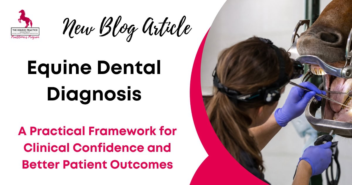
Equine Dental Diagnosis: A Practical Framework for Clinical Confidence and Better Patient Outcomes
If you are an equine veterinarian, chances are you’ve felt that nagging uncertainty when looking inside a horse’s mouth. You’ve finished the odontoplasty, the mouth is clean, but you spot something subtle – a tiny discoloration in a pulp horn, a slightly widened interdental space, or an apex on a radiograph that looks different to the rest.
The internal dialogue begins: “Am I interpreting this correctly? Is this pathology significant or just normal variation? Am I missing something important?”.
This feeling of being stuck is what I call the “equine dentistry treadmill”. We stay busy seeing cases and reducing enamel points, but without a clear framework for equine dental diagnosis, we often fail to move beyond the procedure to true clinical reasoning.
Establishing a definitive diagnosis is the most vital part of our role – it is the difference between being a technician who “floats” and a clinician who treats disease. That diagnostic mindset sits at the heart of modern equine dentistry, where examination, pathology recognition and appropriate diagnostics guide treatment decisions.
The American Association of Equine Practitioners recognises equine dentistry as encompassing the evaluation, diagnosis, prognosis, treatment and prevention of diseases and conditions affecting the teeth and oral cavity
What Equine Dental Diagnosis Actually Is (And Why It Matters)
For too long, general practice dentistry was synonymous with ‘floating’ – the mechanical reduction of sharp enamel points. While preventative odontoplasty is important, equine dental diagnosis is a separate clinical discipline.
So what does that actually mean? It is the deliberate process of transitioning from mere observation to active interpretation and clinical decision-making. It involves:
- Systematic Observation: Utilizing a speculum, focal lighting, and a mirror or oroscope to identify abnormalities across all four cheek tooth arcades (teeth 06-11) and the incisors.
- Pathological Interpretation: Determining if a finding is an acquired pathology, such as periodontal disease or a non-vital pulp, rather than a physiological or anatomical variation.
- Evidence-Based Decision-Making: Using clinical reasoning and advanced diagnostics like equine dental radiography to formulate a treatment plan that is clinically justified.
Because here’s the thing: we are doctors. Our primary objective must be the identification of pathology that impacts patient welfare. Periodontal disease is one of the most common diseases in mammals, yet it is frequently silent. If we aren’t actively looking for it using a structured diagnostic process, we miss the opportunity to intervene before disease becomes irreversible.
Why Most Veterinarians Struggle with Equine Dental Diagnosis
The struggle to establish a confident horse dental diagnosis usually isn’t due to a lack of effort; it’s often due to systemic clinical bottlenecks:
- Suboptimal Diagnostic Environment: Rushing an exam under light sedation or inadequate lighting (like a camping headlamp) is a disaster waiting to happen. You cannot diagnose what you cannot see.
- Over-reliance on Odontoplasty: When the “float” is the goal, we often skip functional assessments. An equine dental assessment should always include a check for lateral molar excursion (EMC) before the speculum goes on, as it can reveal issues with cheek tooth occlusion or the TMJ.
- Uncertainty with Subtle Findings: Differentiating between a healthy infundibulum and a non-vital pulp requires specific visual and tactile skills.
Scientific Note: For pulp assessment, if a pulp explorer or needle sinks into a pigmented area or reveals food material, it represents an exposed, non-vital pulp. Conversely, a “glass-like” sound and feel when scratched indicates the pulp is protected by secondary and tertiary dentine.
A Practical Framework for Equine Dental Diagnosis in Practice
To move past guesswork, I encourage every practitioner to adopt a repeatable equine dentistry workflow:
- Identification: Perform a complete visual and manual check of all teeth. For example, you note a widened gap between two cheek teeth.
- Classification: Is this normal or pathological? A gap is a diastema, which is strongly associated with the initiation of the periodontal disease cascade via bacterial fermentation and inflammatory mediators.
- Clinical Significance: Does it matter clinically? Does the diastema show active food impaction, malodour, or gingival recession?. If yes, intervention is warranted.
- Diagnostic Escalation: Do I need equine oral exam diagnosis support from imaging? Conditions like dental sinusitis, apical infections, or EOTRH cannot be fully assessed without equine dental radiography. When imaging is required, correct equine dental radiography positioning is essential if the resulting images are going to be diagnostically useful.
- Targeted Intervention: Formulate a plan ranging from monitoring to specialized procedures like diastema widening or surgical extraction.
How to Apply This to Real Equine Dental Cases
Case 1: Unilateral Nasal Discharge
This clinical sign should immediately trigger a differential for dental sinusitis secondary to an apical infection of the 08, 09, 10, or 11 maxillary cheek teeth. Radiography is essential here to identify fluid lines.
- One thing to be careful of here: The horse’s head must be perpendicular to the ground to ensure fluid lines appear horizontal on the X-ray for accurate interpretation.
Case 2: The Overlong Tooth
It is common to see a hook or tall tooth and want to reduce it immediately. However, you may be treating the symptom rather than the cause. An overlong tooth is often secondary to a missing opposing tooth or a large diastema. You must assess the opposing arcade to identify the primary horse dental pathology diagnosis.
Case 3: Suspected Non-Vital Pulp
Pulp exposures can be iatrogenic, secondary to high-grade infundibular caries or tooth fractures, or of unknown etiology. A non-vital pulp indicates the tooth has stopped laying down secondary dentine, leading to root blunting and eventually apical infection.
Common Mistakes in Horse Dental Diagnosis (And How to Avoid Them)
- Treating Without Diagnosing: Never float teeth simply because it’s a “routine appointment”. Perform a sedated diagnostic exam first and let the pathology guide the treatment.
- Overcalling vs. Undercalling: Don’t extract every tooth that looks “different”. Maloccluded teeth may be healthy if lateral excursion is functional and there is no food impaction. Conversely, never ignore a closed-valve diastema; it is a serious pathology.
- Inaction Due to Uncertainty: If you find a subtle abnormality, do not just “monitor it” out of clinical discomfort. Use your diagnostic tools – primarily radiography – to get clarity.
What This Means in Practice
Elevating your dentistry service starts with a commitment to equine dental clinical reasoning. By implementing a structured diagnostic framework, focusing on interpreting findings rather than just identifying enamel points, and knowing when to escalate to radiography, you reduce guesswork and improve patient outcomes.
Confidence in this field does not come from more “floating” alone; it comes from a deep understanding of the pathology you are treating and the repeatable process used to manage it.
If you’re finding that you want more structure around this, and a clearer way to approach cases like this in practice, that’s exactly what we go through in more detail inside the training.