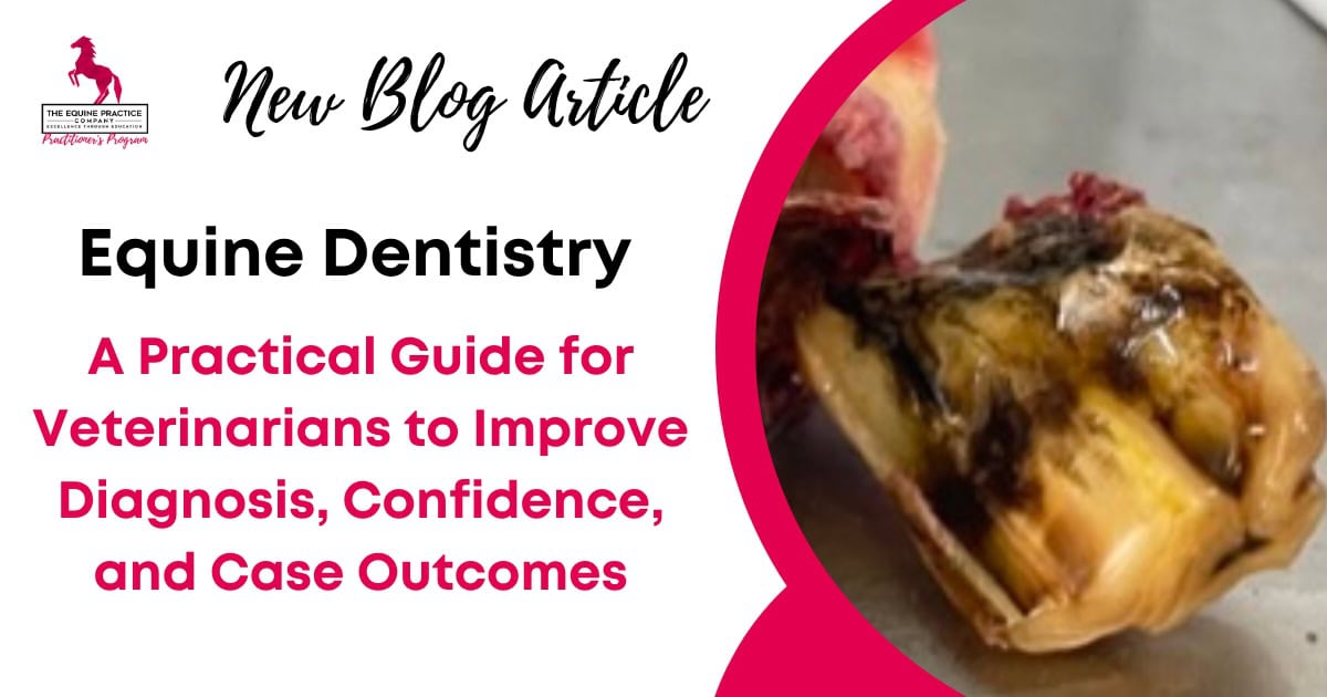
Equine Dentistry: A Practical Guide for Veterinarians to Improve Diagnosis, Confidence, and Case Outcomes
If you’re an equine veterinarian, chances are you’ve felt that nagging uncertainty when it comes to equine dentistry. You’re busy, you’re trying your best, and you’re doing the routine floats. But you might also be wondering:
Am I interpreting this pathology correctly?
Am I missing something important?
Is this simply normal variation or a subtle sign of disease?
I call this the “equine dentistry treadmill.” You’re doing just enough to get by, perhaps using the same gear you’ve had for years, and charging whatever you’ve always charged, without a clear plan to elevate your clinical standard.
The reality is, most of us in this profession were left to piece equine dentistry together on our own. A lecture here, a course there, a few cases, and a lot of second-guessing in between. But the impact of missing pathology, or simply not having a consistent clinical framework, is significant – for our patients and for our own confidence.
This guide is designed to move you off that treadmill. It will bring structure, clarity, and genuine clinical confidence to your equine dentistry service by focusing on the diagnostic discipline, not just the mechanical procedure.
What Equine Dentistry Really Involves
Moving Beyond the Float in Equine Dentistry
Because here’s the thing… equine dentistry is about far more than just floating teeth. If all you’re doing is reducing enamel points, you will be missing a vital part of your role as a diagnostician.
Dentistry is a diagnostic discipline. It requires a clinical mindset, not a technician’s approach. The American Association of Equine Practitioners also defines equine dentistry as encompassing the evaluation, diagnosis, prognosis, treatment and prevention of diseases and conditions affecting the teeth and oral cavity.
It involves a systematic search for pathology that, more often than not, is silent.
So what does that actually mean? It means setting yourself up for success every single time you approach a horse. So many equine practitioners start off on the wrong foot, not because they lack skill, but because they lack the proper setup.
If you don’t have the right sedation, if you have trouble titrating the dose for the individual patient, if your equipment doesn’t allow for a complete oral exam, you’re effectively trying to diagnose in the dark. It’s no wonder so many talented veterinarians walk away from dentistry feeling frustrated – they were never set up to succeed in the first place. You need the right tools and the right knowledge to confidently assess the whole mouth, from the incisors to the last molar, and everything in between.
The Welfare Implications of Missed Dental Pathology in Horses
The simple truth is that horses are masters of disguise. As prey animals, many horses with significant dental disease show few or no obvious outward signs. They often look completely normal. I have seen horses with fractured jaws that haven’t missed a beat with their eating.
When horse owners tell you, “My horse doesn’t need his teeth done because he looks absolutely fine and he’s fat,” you need to recognise that this is a classic misconception that falls on us, as clinicians, to correct.
Welfare implications of missed pathology include:
Chronic pain, particularly with periodontal disease, which can quietly erode quality of life over time.
Dental infections that extend into surrounding structures, leading to conditions like sinusitis and unilateral nasal discharge.
Compounded developmental issues in young horses, where early malocclusions go untreated and worsen as the horse ages.
If you want to see how this approach is applied in real cases, including how to structure your exam and identify pathology early:
The Equine Dental Exam: What Good Actually Looks Like
A high-standard equine dental exam begins before the speculum even goes in.
The Systematic Approach to an Equine Oral Exam
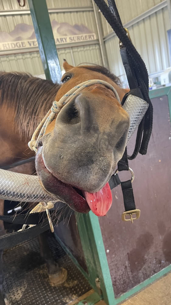
A complete, safe, diagnostic-quality oral exam generally requires adequate sedation. In most cases, you need a sedated, relaxed horse to perform a complete and accurate oral exam. This is essential for safety, but more importantly, for clinical quality.
The external head exam is your starting point. Palpate the temporal muscles for symmetry, palpate down the sides of the cheek for overlong or missing teeth, check for any abnormalities and symmetry of the ventral ramus of each side of the mandible, and assess the incisor and canine teeth. Often, we can identify indicators of a problem before we even open the mouth just from this information.
The full oral exam must be performed using a speculum, a bright focal light source, and a mirror. Modern lighting has dramatically changed our ability to see and diagnose subtle pathology compared to the days of relying on a camping headlight, which simply isn’t strong or focused enough. You need clear vision to identify the subtleties that matter.
Molar Excursion and Functional Assessment
Excursion to molar contact (EMC) is a crucial diagnostic step that is often skipped. With the mouth closed, measure the lateral movement of the jaw by sliding the mandible laterally on both sides. Ideally, there should be a measurable and reasonably symmetrical lateral excursion. Typically, around one to one-and-a-half tooth widths, and the distance should be equidistant from left to right.
If the mouth opens almost straight away, this suggests a problem with the cheek teeth that warrants further investigation. It tells you the horse is unable to perform the full lateral excursion required for their masticatory power stroke. If the separation occurs at a distance greater than this or not at all, it means that the cheek teeth are not in occlusion and may indicate that they have been overfloated.
What Vets Often Skip or Rush
One of the most common mistakes practitioners make is rushing the examination and focusing solely on odontoplasty.
Because here’s the thing… you are there to find and treat disease.
Skipping the external exam, omitting excursion measurements, or failing to properly clean the mouth will inevitably lead to missed pathology. You are unlikely to identify pathology if you are not actively looking for it.
How to Identify Equine Dental Pathology in Horses
Normal vs Abnormal
Understanding what is normal is the prerequisite for identifying pathology. While the anatomy of the horse’s mouth can be complex, the key lies in recognising the patterns that matter.
For example, deciduous teeth only occur on premolar, not molar cheek teeth. This is a small detail, but it saves time and helps you focus your assessment where it actually matters.
Periodontal Disease in Horses
Periodontal disease is one of the most common and underdiagnosed dental diseases in horses, particularly in general practice. With some studies showing that periodontal disease affected over 60% of horses aged 15 years or more, it is the most common disease of the horse, hands down.
It often begins with an abnormal occlusion or a diastema that traps food. That food then undergoes microbial decomposition, leading to inflammation, bacterial overgrowth, and eventually periodontal pocket formation. But it gets worse, vertical and horizontal bone loss, destruction of the periodontal ligament and eventually endodontic involvement leading to tooth loss or shedding of the tooth. This is a painful problem.
Closed valve diastemas are particularly problematic. The occlusal surface appears tight, but with every chew, food is drawn deeper into the space with no way to escape.
Treatment in these cases often involves removing the impacted feed, reducing malocclusions on the opposing surface and widening the diastema to allow food to move freely, reducing ongoing disease progression.
Pulp Exposure vs Non-Vital Pulp
And this is where it gets interesting…
Correctly identifying a non-vital pulp is one of the most important diagnostic skills in equine dentistry.
A vital pulp feels smooth and glass-like when probed, protected by secondary and tertiary dentine. A non-vital pulp that is open to the oral environment, on the other hand, allows penetration, often appears discoloured, and may contain food material.
Once the pulp becomes non-vital, part or the tooth or even the entire tooth (the endodontic system) is ‘dead’, and production of dentine ceases. Because the periodontal system is a totally different system, the tooth will still continue to erupt. This can lead to apical infections and may ultimately even require extraction.
Equine Dental Radiography: When and Why It Changes Everything
Of all the areas dentistry veterinarians struggle with, equine dental radiography is consistently at the top.
And that’s understandable.
It’s technically challenging, and interpretation can be difficult without the right framework.
When to Take Equine Dental Radiographs
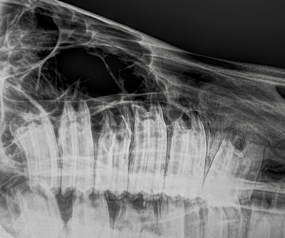
Radiographs are indicated whenever significant dental or sinus pathology is suspected. Radiographs should be considered in cases involving suspected endodontic disease, deep periodontal pockets, some malocclusions, sinusitis, facial swelling, draining tracts, trauma, or fractured teeth.
The Importance of Correct Positioning
If you don’t remember anything else, remember this: the horse’s head must be perpendicular to the ground when taking lateral sinus views. The bisecting angle technique is your best friend, you don’t need to memorise the angles.
If your positioning is off, your interpretation will be too.
Radiography is what transforms equine dentistry from assumption to a more evidence-based diagnosis.
What Most Veterinary Dentists Miss in Equine Dentistry
Many veterinary dentists who feel stuck are often missing the same few key elements.
They underestimate the opportunity already sitting within their existing client base. They assume cases are routine when they are not. And most importantly, they lack a structured approach to assessment and decision-making.
Dentistry becomes clearer and more rewarding when you have a repeatable system.
A Practical Framework for Better Equine Dentistry in Practice
If you want to improve your outcomes and confidence, the shift is simple – but not easy.
Perform a thorough and systematic examination every time. Focus on identifying pathology. Communicate clearly with clients. Recommend appropriate next steps.
This is not about doing more work. It is about doing better work.
Common Mistakes in Equine Dentistry (And How to Avoid Them)
The most common mistakes I see are not due to lack of knowledge, but lack of structure.
- Rushing the exam increases the risk of missed disease.
- Skipping appropriate diagnostics increases clinical uncertainty.
- Undervaluing services limits growth.
- Poor communication reduces client uptake.
Each of these is fixable with the right framework.
What This Means in Practice
To improve your equine dentistry service:
- Perform a complete oral exam every time.
- Focus on diagnosis, not just treatment.
- Use radiographs when indicated.
- Communicate clearly with clients.
- Recognise dentistry as a significant opportunity within your practice.
You can also explore our latest equine dentistry articles and clinical resources for veterinarians.
FAQs About Equine Dentistry
How often should horses have a dental exam?
Most horses should have a dental examination at least once per year, although this should be tailored to the individual.
What is the most common dental disease in horses?
Periodontal disease is the most common and often the most underdiagnosed condition seen in equine dentistry.
When should equine dental radiographs be taken?
Radiographs should be taken whenever pathology is suspected, particularly in cases involving sinus disease, swelling, or abnormal clinical findings.
What is the difference between a routine float and a proper dental exam?
A routine float focuses on reducing enamel points, whereas a proper dental exam is a full diagnostic assessment of the oral cavity, including identification of pathology and treatment planning.
Do you have a program suitable for equine veterinarians who wish to become a specialist?
A routine float focuses on reducing enamel points, whereas a proper dental exam is a full diagnostic assessment of the oral cavity, including identification of pathology and treatment planning.
Do you offer an online equine dentistry program for veterinarians who want to develop advanced dental skills?
Yes. The Equine Practice Company’s Equine Dentistry Program is designed for equine veterinarians who want to build greater confidence and capability in equine dentistry, including systematic dental examinations, pathology recognition, diagnosis, radiography, treatment planning and more advanced case management. The focus is on helping veterinarians move beyond routine floating and develop a more diagnostic, clinically structured approach to dentistry. This matches the clinical approach outlined throughout this guide.
Closing
If you’re finding that you want more structure around this, and a clearer way to approach cases like this in practice, that’s exactly what we go through in more detail inside the training.
It’s about building confidence through clarity, and giving you a repeatable system you can rely on in every case.