Free Practitioner’s Program Training Videos

Podiatry

Measurements of the Hoof: Bone Angle

There are many measurements that are performed when assessing podiatry radiographs, such as bone angle and understanding how that may affect the external look of hooves.

X-Ray Principles 4: kVp and mAs for Perfect Images

There are 4 main X-ray principles, one of them being kVp and mAs which affects contrast and exposure. See how an overexposed image can affect a vet’s view as well as how to create the perfect image.

Veterinary & Podiatry Radiographs

Understand the difference between veterinary and podiatry radiographs, along with the techniques for each.

Two Block Technique

Jen Lugton explains the two block technique when performing radiographs on hooves to ensure accurate images.

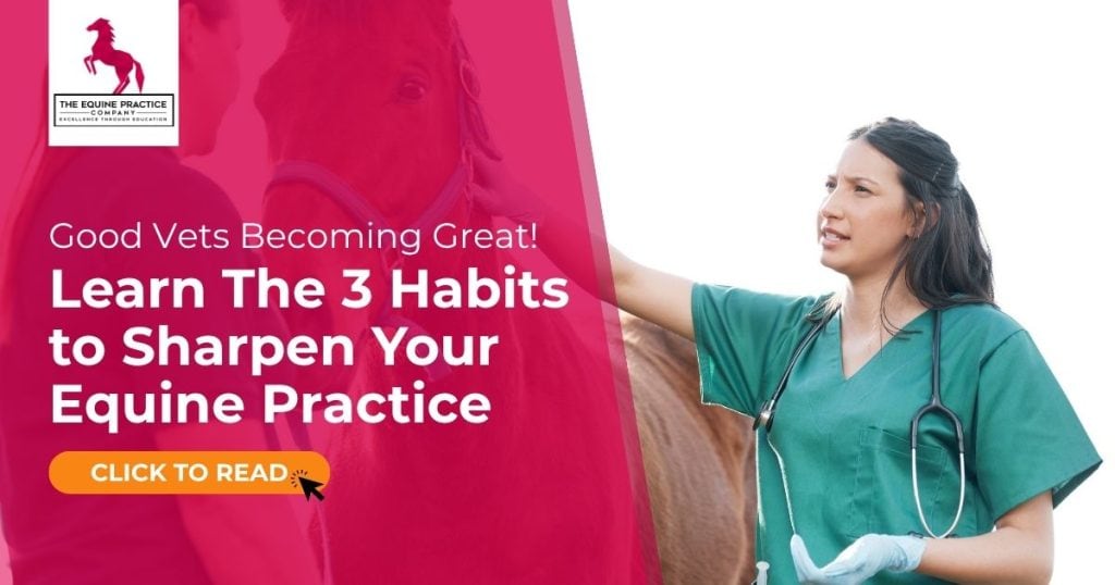
How Good Vets Become Great: 3 Habits to Sharpen Your Equine Practice

I’ve asked myself this question more times than I can count, especially in my early years of practice. Would I trust myself with my own horse? On a good day? Maybe.On a bad day? I’m…

X-Ray Principles 2 &3: Magnification & Reducing Distortion to Improve Accuracy

Jen Lugton explains more X-Ray principles to ensure a great radiographic image is created by setting magnification, reducing distortion and improving accuracy.

X-Ray Principles 1: Focal Beam Orientation

In this video, Jen Lugton explains the first X-ray principle and how orientation of focal beams can affect radiographs

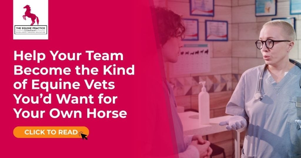
How Great Clinics Are Built: 3 Habits of High-Performing Teams

Ever wondered what makes some equine clinics thrive while others plateau? These 3 clinic-wide habits can sharpen clinical standards, improve case outcomes, and boost team retention – fast.

Fundamentals of Equine Podiatry Imaging: Improving Diagnostic Accuracy in the Field

High-quality podiatry radiographs are essential for accurately assessing hoof balance, diagnosing pathology, and guiding farriery decisions. Consistent technique, correct positioning, and a solid understanding of radiographic principles are what separate routine images from genuinely diagnostic ones. The videos on this page provide a practical framework for producing reliable, repeatable hoof radiographs in everyday practice.

  • Understanding Key Hoof Measurements and Why They Matter
    Bone angle, palmar angle, sole depth, joint alignment, and dorsal wall relationships are critical markers when evaluating hoof balance. Small variations in these measurements can lead to significant biomechanical consequences. Accurate imaging allows veterinarians to assess conformation, detect early pathology, and communicate effectively with farriers when planning corrective trimming or shoeing.
  • Radiographic Principles: kVp, mAs, Magnification and Distortion
    Clear, diagnostic images depend on controlling contrast, exposure, and beam positioning. Knowing how adjustments in kVp and mAs influence image quality helps practitioners avoid overexposure, underexposure, and loss of detail. Equally important is managing magnification and minimising distortion – both common pitfalls in hoof radiography that can misrepresent angles and distances.
  • Veterinary vs Podiatry Radiographs
    Although the techniques share similarities, podiatry-specific radiographs require unique positioning, targeted views, and a high emphasis on detail. These images must reliably highlight the distal phalanx, laminar zone, joint spaces, and soft tissue outlines. Understanding the differences between standard veterinary radiographs and podiatry-focused techniques ensures the practitioner captures the most clinically relevant information.
  • Two-Block Technique for Accurate Positioning
    Consistent, level positioning is the foundation of high-quality hoof imaging. The two-block method stabilises the foot, aligns the distal limb, and helps ensure the beam passes through the correct anatomical landmarks. This reduces variation between images and allows for confident comparison over time or between limbs.
  • Focal Beam Orientation and Its Role in Image Accuracy
    Even subtle changes in beam orientation can significantly alter how anatomical structures appear. Properly aligning the beam with the hoof capsule and skeletal landmarks ensures that measurements taken from the radiograph reflect true anatomy rather than projection error. Mastery of beam direction is one of the most effective ways to improve diagnostic accuracy.
  • Recognising and Minimising Common Radiograph Errors
    Foot not placed correctly on the plate, improper angulation, inconsistent height, and rotation are among the most common causes of poor podiatry images. Becoming familiar with these patterns helps veterinarians identify errors immediately and correct them before they impact diagnosis or farriery planning.