Equine Hock Radiographs: A Systematic Approach to Acquisition and Interpretation
Equine hock radiographs can reveal substantial abnormalities, subtle changes or apparently normal anatomy in a horse with tarsal region pain. Interpreting them requires more than identifying new bone or a narrow joint space.
The images must be of diagnostic quality, the anatomy must be understood, and the findings must be considered alongside the clinical examination and responses to diagnostic anaesthesia.
In her recent webinar, Dr Sue Dyson emphasised two recurring problems: inadequate image acquisition and overinterpretation of minor radiographic abnormalities. Both can lead the investigation away from the actual source of pain.
Her approach begins with positioning the horse appropriately for optimal image quality and the acquisition of a complete set of projections, followed by systematic assessment of every image. The final question is whether the abnormalities identified explain the horse’s clinical presentation.
Start with positioning and image quality
For routine assessment, position the metatarsal region vertically and account for the horse’s natural hindlimb orientation. Many horses stand with their hind feet turned out, which influences the beam direction needed for lateromedial and dorsoplantar projections.
The distal tarsal joint spaces are not horizontal. They slope distally from lateral to medial and, to a lesser extent, from dorsal to plantar. The x-ray beam therefore needs to follow the relevant joint orientation if the spaces are to be assessed reliably.
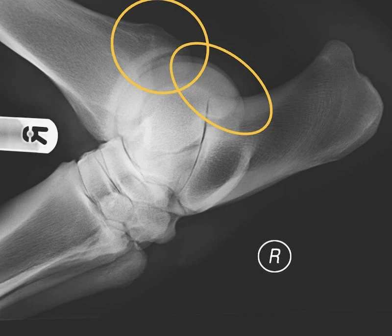
For the lateromedial projection, direct the x-ray beam tangential to the heel bulbs, together with appropriate adjustment for the slope of the distal joints. Treat positioning as something to assess in the individual horse rather than relying on a fixed angle for every examination.
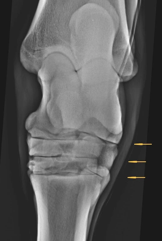
If the metatarsus is not vertical or the beam angle is inappropriate, overlapping bone margins can obscure the joint spaces. A poorly demonstrated space cannot be interpreted confidently as normal or narrowed and subchondral bone changes may be missed..
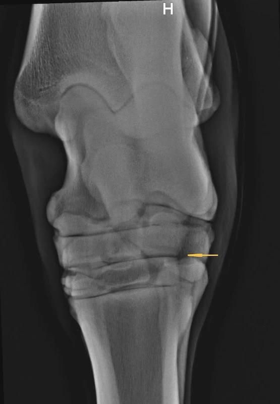
If several joint spaces appear reduced in width on the same side of a dorsoplantar image it is probably the result of positioning. Before concluding that multiple joints are affected, reacquire the image but alter the angle of the x-ray beam.
Acquire at least four routine projections
A minimum of four projections should be acquired for routine radiographic evaluation of the hock:
- Lateromedial.
- Dorsoplantar.
- Dorsolateral–plantaromedial oblique.
- Dorsomedial–plantarolateral oblique.
Each provides different information, and an abnormality may be apparent on only one projection. A convincing finding on one image should therefore prompt careful assessment of the remaining series, while an apparently unremarkable image should not replace the other views.
Joint space narrowing is usually best assessed on a well-positioned dorsoplantar projection. Subchondral radiolucent areas in the centrodistal and tarsometatarsal joints are often better demonstrated on the dorsolateral–plantaromedial oblique projection.
The routine series is a starting point. Additional projections should be selected according to the clinical findings and the anatomy requiring closer evaluation. For example, a flexed lateromedial projection can improve assessment of the proximal portions of the trochleas of the talus and the caudodistal aspect of the tibia.
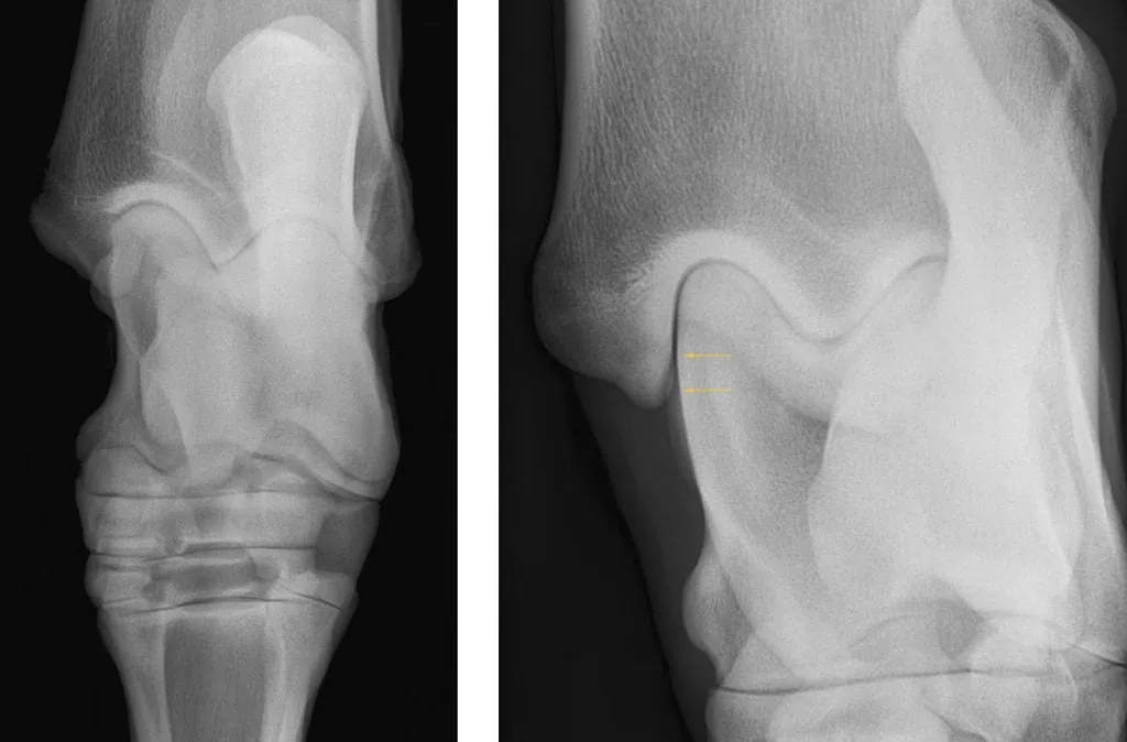
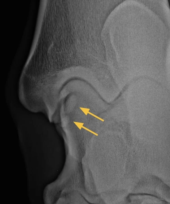
Recognising superimposition is also essential. On a true dorsoplantar projection, parts of the proximal talus and the axial aspect of the medial malleolus overlap.
When that region requires investigation,for example in a horse with tarsocrural joint effusion, an additional dorsal 15°lateral-plantaromedial oblique projection may reveal fragments on the axial aspect of the medial malleolus.
For another example of how positioning and projection choice affect interpretation, see our guide to evaluating the palmar compact bone on navicular radiographs.
Use the same interpretation sequence every time
A systematic review reduces the risk of stopping at the first conspicuous abnormality. Dr. Dyson’s assessment includes the soft tissues, bone margins and shape, subchondral bone, trabecular architecture, joint spaces and interosseous spaces.
Begin by checking whether the image is suitable for evaluating the structures of interest. Then work through the following questions:
| Structure | Questions to ask |
| Soft tissues | Is there swelling or a change in the expected outline? |
| Bone margins and shape | Are the contours normal? Is there new bone, a defect or altered shape? |
| Subchondral bone | Are the subchondral bone plate thickness and contour regular? Are there areas of increased thickness or radiolucency? |
| Trabecular bone | Is the normal architecture preserved, or is there a regional change? |
| Joint spaces | Can they be assessed adequately, and is their width regular? |
| Interosseous spaces | Are they clearly defined, with an expected outline and surrounding bone appearance? |
The anatomy of the hock region is highly complex with multiple bones and soft tissues. Knowledge of soft tissue attachments helps interpret new bone at the margins of the tarsus. An osseous prominence needs to be assessed in relation to both the adjacent joint and nearby ligament or tendon attachments.
Anatomical specimens and good reference images are useful when an appearance is unfamiliar. Understanding how bones overlap in different projections can resolve uncertainty that image inspection alone may not.
Distinguish normal anatomy and projection effects from disease
Not every radiolucent line represents a fracture, and not every rounded lucency represents a cyst-like lesion. Normal osseous canals and superimposed bone margins can create appearances that invite overinterpretation.
A Mach line is one example: a lucent line which is the result of one bone being partially superimposed over another can resemble a fracture line. Recognising the underlying anatomy and reviewing the other projections are necessary before assigning pathological significance.
The same discipline applies when evaluating serial radiographs. Differences in limb position or beam direction may alter the apparent size and shape of a feature. Before concluding that a lesion has progressed, assess whether the images are sufficiently comparable.
The practical aim is to describe what is present accurately, then determine its significance. A familiar-looking abnormality should not bypass that process.
An osseous spur is not a diagnosis of pain
Spurs on the dorsoproximal aspect of the third metatarsal bone illustrate why radiographic findings need clinical context.
In a retrospective study of 455 horses, Fairburn, Dyson and Murray found these spurs in 25% of horses. Their frequency did not differ significantly between lame and nonlame horses. Spurs were associated with the severity of radiographic abnormalities of the centrodistal joint, but an isolated spur could be incidental. Radiographic differentiation between an osteophyte and an enthesophyte at the insertion of tibialis cranialis or fibularis tertius or the dorsal tarsal ligament was often difficult. [1]
These findings support examining the adjacent joints carefully without assuming that the spur explains the lameness. Consider the entire radiographic series and the results of the clinical investigation, including diagnostic anaesthesia.
For an individual horse, the presence of a spur alone does not establish whether it is painful or predict how the appearance will change over time.
For a related discussion using navicular examples, see our guide to interpreting radiographic findings in clinical context.
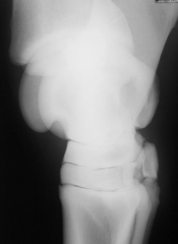
Time zero – there is a small spur on the dorsoproximal aspect of the third metatarsal bone. Identical findings were seen in both hindlimbs. Right hindlimb lameness was abolished by intra-articular anaesthesia of the tarsometatarsal joint.
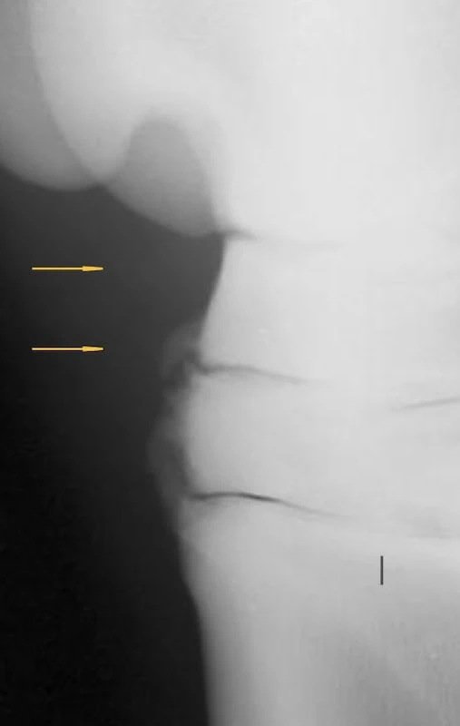
Four years later. Periarticular osteophyte formation has progressed in the right hindlimb. The left hindlimb was unchanged.
Include the interosseous ligament regions
The interosseous spaces deserve deliberate assessment rather than being overlooked while attention is directed towards periarticular new bone.
In their study of 700 horses and ponies, Skelly-Smith, Ireland and Dyson described the normal centrodistal interosseous space as an oval or circular radiolucency with a regular rim of bone proximally and distally.
Rim thickness varied between horses. Abnormalities of this region were associated with radiographic centrodistal osteoarthritis, but either finding could occur without distal tarsal joint pain. [2]
The webinar highlighted mineralisation within the region and changes in the surrounding bone as features to assess. These observations form part of the overall examination; their presence should not be treated as proof that this is the painful structure.
Assess the pattern of distal tarsal joint changes
Distal tarsal osteoarthritis can involve periarticular new bone, altered joint space width and changes in the subchondral bone. The combination and distribution of these findings matter.
Resorptive subchondral bone changes are a particular cause for concern. These warrant careful attention when evaluating a horse with lameness, even if an intra-articular block has not produced the expected improvement.
Intra-articular anaesthesia may not affect subchondral bone pain, however pain causing lameness should be abolished by perineural anaesthesia of the tibial and fibular nerves.
However, the overall relationship between radiographic appearance and clinical signs is not straightforward.
In a retrospective study of 91 horses with small tarsal joint osteoarthritis and positive intra-articular anaesthesia, Byam-Cook and Singer found no association between radiographic findings and the duration or degree of lameness, or the response to diagnostic anaesthesia. [3]
It is therefore inappropriate to translate the amount of visible change directly into a pain score. Equally, mild radiographic findings should not bring an investigation to an end when they do not adequately explain the clinical presentation.
Progression is also variable. Contrary to old literature not every affected distal tarsal joint will proceed to complete fusion with subsequent relief of pain and lameness. Progression is unpredictable.
The distinction between an imaging abnormality and its clinical significance is also explored in our review of radiographic abnormalities in performance horses, which examines findings in the thoracolumbar spine.
Interpret diagnostic anaesthesia alongside the images
Diagnostic anaesthesia is central to determining the relevance of radiographic abnormalities, but its limitations must be understood.
There is overlap between the effects of nerve blocks used in the distal tarsal and proximal metatarsal region. Intra-articular anaesthesia of the tarsometatarsal joint can influence pain associated with the proximal suspensory region.
Conversely, anaesthesia of the deep branch of the lateral plantar nerve can influence distal tarsal joint pain.
An improvement after either procedure therefore needs interpretation within the wider investigation, with grading of the improvement after each block. It does not, by itself, provide an anatomically exclusive diagnosis.
Comprehensive radiographic and ultrasonographic evaluations of the hock and proximal metatarsal regions are required.
A negative response to intra-articular anaesthesia does not necessarily exclude tarsal region pain. Pain with an important subchondral bone component may not be abolished by intra-articular anaesthesia.
In selected investigations, tibial and fibular nerve blocks can contribute to further localisation, with their responses considered alongside the other findings.
This is particularly relevant when the severity of lameness and the apparent radiographic explanation do not agree. The investigation should continue rather than assigning the pain to the only visible abnormality.
Recognise when radiography is insufficient
Radiography provides valuable information about bone, but limited information about soft tissues. Dr. Dyson emphasises that soft tissue injuries are an important part of tarsal region disease and that ultrasonography is complementary to radiography.
Bone, joint and soft tissue injuries may co-exist. More than one site of pathology can contribute to pain and lameness.
Radiographs may also underestimate the extent of some joint and bone lesions. An absence of convincing abnormalities does not exclude tarsal region pathology, including some forms of osteoarthritis, bone injury or ligament injury.
When diagnostic anaesthesia has localised pain but radiography and ultrasonography have not provided an adequate explanation, advanced imaging may be appropriate using either CT or MRI.
The choice of which to use will depend to some extent on what facilities are available, bearing in mind that there are differences in quality of fan beam and cone beam CT images for both osseous and soft-tissue structures, and those acquired standing versus in recumbency under general anaesthesia.
Likewise low-field and high-field MR images are different in quality and the information that can be acquired.
Reliable interpretation of equine hock radiographs rests on this continued integration of findings: diagnostic-quality images, a consistent anatomical review, and a clinical investigation that tests whether the visible changes account for the horse’s pain.
Sources
Primary source: Dr Sue Dyson’s September 2026 webinar on optimising hock radiographs and image interpretation, Other sources include:
- Fairburn A, Dyson S, Murray R. Clinical significance of osseous spurs on the dorsoproximal aspect of the third metatarsal bone. Equine Veterinary Journal. 2010;42(7):591–599. Study abstract and publication details.
- Skelly-Smith E, Ireland J, Dyson S. The centrodistal joint interosseous ligament region in the tarsus of the horse: Normal appearance, abnormalities and possible association with other tarsal lesions, including osteoarthritis. Equine Veterinary Journal. 2016;48(4):457–465. Study abstract and publication details.
- Byam-Cook KL, Singer ER. Is there a relationship between clinical presentation, diagnostic and radiographic findings and outcome in horses with osteoarthritis of the small tarsal joints? Equine Veterinary Journal. 2009;41(2):118–123. Study abstract and publication details.